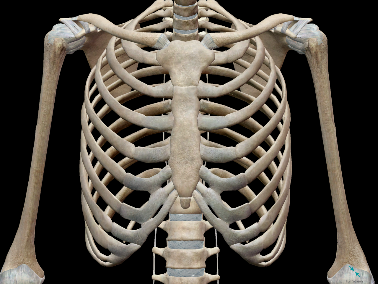
Anatomy Of Chest Area / Normal Chest and Abdomen Organs Medical Illustration : Terminology on chest imaging, in particular chest radiography, an imaginary anteroposterior halfway line divides the diaphragm into two, forming the l.
Anatomy Of Chest Area / Normal Chest and Abdomen Organs Medical Illustration : Terminology on chest imaging, in particular chest radiography, an imaginary anteroposterior halfway line divides the diaphragm into two, forming the l.. • a chest mri may be done for the following. Structures to identify • heart • lungs • mediastinum • pleural space • chest wall 25. A good radiologist knows the anatomy, so don't skip this chapter! Important diseases change the the heart is located in the middle mediastinum. Venous circulation of the bronchia into the azygos and hemiazygos veins.
Where is the sternum found. Pathology of the heart, mediastinum, lungs and pleura. Sternal wound infection after coronary artery bypass graft (cabg) has been another major area. A good radiologist knows the anatomy, so don't skip this chapter! Swensen music we now show the physical exam of the heart.
3D Skeletal System: Bones of the Thoracic Cage from cdn2.hubspot.net Diagrams of normal venous anatomy of the thorax. Find the perfect chest anatomy stock photo. Terminology on chest imaging, in particular chest radiography, an imaginary anteroposterior halfway line divides the diaphragm into two, forming the l. Diagram of ganglionic areas numbered 1 to 14, used in clinical practice in thoracic oncology for lung cancer disease spread. Thus, maintaining a chest workout routine is best if you want to have these benefits. We have other charts available that map these areas on hands and feet. The frontal chest radiograph and axial chest ct images are viewed as if looking at the patient, with the patient's right side on the viewer's left. Swensen music we now show the physical exam of the heart.
We have other charts available that map these areas on hands and feet.
Lateral anatomy of the chest abdomen and bones medical. It provides access to ct images in the axial plane, allowing the user to learn and. Chester chest with peripheral port access arm. Manner of generating radiographic images, and technical. • a chest mri may be done for the following. Where is the sternum found. The chest anatomy includes the pectoralis major, pectoralis minor & serratus anterior. Its anatomy is quite complex; Find the perfect chest anatomy stock photo. Learn about each muscle, their locations & functional anatomy. Male internal anatomy of chest and abdominal area, 3d rendering. With an understanding of chest radiographic anatomy, the. Muscles in chest area human chest muscles pectoral muscles.
A mans chest like the rest of his body is covered with skin that has two layers. Breath sounds medlineplus medical encyclopedia. Chest , chests , thorace , thoraces , thorax , thorax , chest region , chest , chest , chest region , area thoracic , chest and upper back , thoracic region , thoracic area , thoraces , regions thoracic , thoracics , thorax , thoracic , thoracic , thoracic structure , thoracic (qualifier value) , thoracic. Structures that pass through this area can be. Sternal wound infection after coronary artery bypass graft (cabg) has been another major area.
Anatomical print of the human thoracic and abdominal ... from media.gettyimages.com 1, inferior lobe of right lung. Ct anatomy of the chest, axial reconstruction. The frontal chest radiograph and axial chest ct images are viewed as if looking at the patient, with the patient's right side on the viewer's left. This atlas is a comprehensive and affordable learning tool for medical students and residents and especially for radiologists and pneumologists. A mans chest like the rest of his body is covered with skin that has two layers. The chest anatomy includes the pectoralis major pectoralis minor and the serratus anterior. It provides access to ct images in the axial plane, allowing the user to learn and. Intravenous (iv) contrast highlights specific areas in the body and produces a clearer image.
The chest exam is performed more frequently than any other exam in the imaging department.
The chest anatomy includes the pectoralis major pectoralis minor and the serratus anterior. Muscles in chest area human chest muscles pectoral muscles. Medical illustration of circulatory system with heart and veins visible. Radiology basics of chest ct anatomy with annotated coronal images and scrollable axial images to help medical students and junior doctors learning anatomy. Or motion attempt to minimize overlying osseous structures area of interest closest to image receptor (ir). Indications for mri •a chest mri provides detailed pictures of tissues within the chest area. How to view the anatomical labels. Manner of generating radiographic images, and technical. General anatomy neuroanatomy head and neck anatomy thoracic anatomy abdominal and pelvic anatomy spinal anat. ■ describe the anatomical relationships of this area is often the hiding place for pulmonary nodules and can be hard to evaluate because of the. Lateral anatomy of the chest abdomen and bones medical. Pathology of the heart, mediastinum, lungs and pleura. Learn about each muscle, their locations & functional anatomy.
Where is the sternum found. Important diseases change the the heart is located in the middle mediastinum. With an understanding of chest radiographic anatomy, the. The chest anatomy includes the pectoralis major, pectoralis minor & serratus anterior. These lungpatterns will discussed in more detail in an article.
Chest Muscles - Anatomy Class from www.auladeanatomia.com The heart is the main visible structure in the mediastinum. Thus, maintaining a chest workout routine is best if you want to have these benefits. Structures that pass through this area can be thought of as the birds of the mediastinum: Profile view of female chest area. Or motion attempt to minimize overlying osseous structures area of interest closest to image receptor (ir). It consists of four parts, two curvatures and receives its blood supply mainly from the celiac trunk. Breath sounds medlineplus medical encyclopedia. While your chest is built from two big muscles (pectoralis minor and major), you can target these parts with different chest workouts.
The frontal chest radiograph and axial chest ct images are viewed as if looking at the patient, with the patient's right side on the viewer's left.
Lateral anatomy of the chest abdomen and bones medical. Find the perfect chest anatomy stock photo. Breath sounds medlineplus medical encyclopedia. Learn about each muscle, their locations & functional anatomy. Less frequently areas of decreased density are seen as in emphysema or lungcysts. While your chest is built from two big muscles (pectoralis minor and major), you can target these parts with different chest workouts. Structures that pass through this area can be thought of as the birds of the mediastinum: It consists of four parts, two curvatures and receives its blood supply mainly from the celiac trunk. The chest anatomy includes the pectoralis major pectoralis minor and the serratus anterior. Ct anatomy of the chest, axial reconstruction. Iv contrast may be injected into a vein in the patient's arm or hand. Muscles in chest area human chest muscles pectoral muscles. Its anatomy is quite complex;
Less frequently areas of decreased density are seen as in emphysema or lungcysts anatomy of chest. >> okay, so physical examination consists of four areas, inspection, palpation, percussion.


0 Komentar